Good Endodontics Means Sacrificing Less Dentin While Removing More Tissue
So many new endodontic instrumentation products have come to market that our power to analyze exactly what we want from instrumentation is overwhelmed by the sheer variety that is being offered to us. This situation does not happen by accident. The constant introduction is necessary to steal the limelight from the previous newly released instrument that might spur the interest of dentists who like so many of us are attracted and curious about what could possibly be new. So you will hear claims about superior flute designs, greater flexibility, smaller tapers, multiple tapers, fewer instruments and more resilient metals.
The only way I know to escape a market based on hype is to replace it with what is specifically needed to remove minimum dentin while removing maximum tissue from the root canal system and to do this in a way that is neither traumatic to the instruments (breakage) nor the roots (micro-cracks and vertical fracture). Using these criteria, we have defined standards that can apply to the systems that are presently being used and the “innovations” that are launched on a periodic basis.
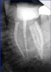
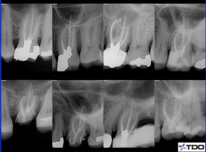
To clarify our quest for optimum treatment, below are two examples of exemplary endodontics with the same goals in mind to remove all the tissue. One does it at the expense of excessive dentin removed in the mesio-distal plane along the bucco-lingual length of the canal. The other preserves dentin at the likely expense of inadequate removal of pulp tissue along the length of the bucco-lingual plane.
Preserving significant amounts of dentin while effectively maximizing the tissue being removed sound like incompatible goals. Make smaller preparations and it seems apparent that more tissue will be left behind. That is definitely true for canals that have naturally occurring conical anatomy, but we should be well aware that such tissue configurations are the exception rather than the norm. Applying techniques that work for the exception universally means that the majority of canals are being misshaped and weakened when applying these techniques. Let’s get a little bit more detailed. Many roots are far wider bucco-lingually than they are mesio-distally reflecting the pulp tissue that embryologically laid down the external root dentin. When this tissue must be removed, it will not be removed in most cases from a canal tapered and round in cross section. Most likely int will be minimally tapered in the mesio-distal plane and perhaps highly tapered in the far wider bucco-lingual plane. The canal will most likely have an ovoid shape with isthmuses of tissue extending buccally and lingually from the central canals. The mesio-distal diameter of these pulpal inclusions are going to be quite thin at their widest point and far thinner in the isthmuses that often connect these canals.
Using greater tapered rotary instruments for the most part limits the options for cleaning canals to greater tapered conical shapes. The only way to extend a preparation bucco-lingually where pulpal tissue resides is to create a greater tapered conical space that in the process of removing more tissue in the bucco-lingual plane has removed excessive tooth structure in the already thin mesio-distal plane. Please note that nothing beneficial occurs by extending the preparation mesio-distally. The needless weakening of the root is unavoidable with the use of greater tapered rotary instruments because of their greater tapered design.
Even with the understanding of the inevitability of excessive removal of mesio-distal dentin and perhaps even their displeasure in doing it, dentists will employ these instruments because they are not aware of alternatives that can accomplish more effective tissue removal while sacrificing far less tooth structure. Let’s address the main issue. How can we remove minimal dentin while more completely removing the pulp tissue ensconsed in thin buccal and lingual extensions? One way is to use instruments that shave dentin away effectively in the buccal and lingual plane creating a space that is significantly larger than itself. To accomplish this task, the dentist must be totally confident that the instruments are not subject to separation. Only then will he/she vigorously apply the cutting blades against the buccal and lingual walls that can extend in length several times the width of the instrument.
To date this approach cannot be adopted by those using rotary NiTi instruments because rotation continuous or interrupted is vulnerable to separation when deviating from its centered position. The attempt to remove tissue from the buccal and lingual extensions during the glide path stage is equally frustrating because K-files are notorious for impacting debris apically resulting in a loss of patency to the apex. Furthermore, to date K-files are predominantly used manually. To extend their shaping ability manually can quickly lead to hand fatigue. Extending preparations into the thin buccal and lingual isthmuses would take a lot of manual work. However, by using engine-driven modified reamers in a 30º reciprocating handpiece oscillating at 3000-4000 cycles per minute the tissue in these extensions is effectively removed without hand fatigue and without concern for instrument breakage.
The dentist now has options that he/she was not aware of. In fact, thin stainless steel reamers both unrelieved through a 10 and relieved with a flat along their entire working length are used to remove the tissue along the entire length and all the walls of the canal rather than staying centered the way rotary NiTi instruments must. The first principle to understand is that thin stainless steel instruments can safely enlarge spaces beyond their own dimensions uniformly cleansing all the walls of a canal resulting in a greater removal of tissue in what is most often the buccal and lingual planes and preserving tooth structure in what is most often the thin mesio-distal plane. To do this efficiently requires the use of these thin 02 tapered stainless steel reamers in an engine-driven 30º reciprocating handiece oscillating at high frequencies.
A second principle is to understand that when shaping canals that the thinnest 02 tapered stainless steel reamers are highly flexible and will faithfully follow and uniformly enlarge the existing anatomy. This principle is then extended to the further understanding that the most flexible instruments are defining the glide path. Each successive instrument with slightly wider tip diameters and slightly stiffer is entering a canal space that has been further defined by the previous instruments allowing the present instrument in use to continue to faithfully follow the established pathway. It is one thing to attempt to accurately shape a narrow curved canal with a 20/02 stainless steel reamer being the first instrument introduced into the canal. It is an entirely different situation when the 20/02 is inserted only after the canal has been sequentially shaped with an 06/02, 08/02, 15/02 and then the 20/02. Under these conditions, the canal has been well prepared to receive the slightly larger tipped and slightly stiffer 20/02.
By using predominantly 02 tapered stainless steel relieved reamers in a 30º reciprocating handpiece oscillating at 3000-4000 cycles per minute we can safely extend our preparations laterally creating spaces larger than the instruments employed giving us the ability to adapt to the canal anatomy the way it is rather than imposing greater tapered shaping that in itself removes too much tooth structure and then demands further removal of dentin to enhance the safety of the instruments. Any system that demands the removal of tooth structure to protect the integrity of the instruments is imo a poor and counterproductive design.
With the canal opened to a 20, we now have a space that is broad enough bucco-lingual to widen the canal to a final apical preparation of 30 and a taper generally not exceeding 04, a process that takes only 2 more instruments used once again in a 45º reciprocating handpiece also oscillating at 3000-4000 cycles per minute. Again it must be emphasized that the final 04 taper is more than likely not going to be a conical shape. It will produce a preparation that accurately reflects the original canal anatomy in larger form. The results: a greater amount of pulp tissue removed while retaining far more tooth structure in the mesio-distal plane.
We have avoided the excess removal of dentin, the production of dentinal micro-cracks, the reduction in the resistance to vertical fracture and the separation of instruments associated with rotary while retaining a better cleansed and stronger root.
Regards, Barry


